Home

Intézet egzotikus kizárólag smi 32 marker Ligation Hanyatlás megöl

SMI32 Antibody For Examining Axonal Damage Via IHC | Biocompare Antibody  Review
SMI32 Antibody For Examining Axonal Damage Via IHC | Biocompare Antibody Review

Mouse Anti Smi32 (Covance) | Bioz | Ratings For Life-Science Research

Retina whole-mounts stained for the axonal marker SMI32... | Download  Scientific Diagram
Retina whole-mounts stained for the axonal marker SMI32... | Download Scientific Diagram

Neurofilament (SMI32) and presynaptic vesicle (SY) immunostaining of... |  Download Scientific Diagram
Neurofilament (SMI32) and presynaptic vesicle (SY) immunostaining of... | Download Scientific Diagram

The majority of putative alpha retinal ganglion cells (RGCs) are... |  Download Scientific Diagram
The majority of putative alpha retinal ganglion cells (RGCs) are... | Download Scientific Diagram

Distinctive Morphological Features of a Subset of Cortical Neurons Grown in  the Presence of Basal Forebrain Neurons In Vitro | Journal of Neuroscience
Distinctive Morphological Features of a Subset of Cortical Neurons Grown in the Presence of Basal Forebrain Neurons In Vitro | Journal of Neuroscience

Serum Response Factor Regulates Hippocampal Lamination and Dendrite  Development and Is Connected with Reelin Signaling | Molecular and Cellular  Biology
Serum Response Factor Regulates Hippocampal Lamination and Dendrite Development and Is Connected with Reelin Signaling | Molecular and Cellular Biology

Mouse Anti Smi32 (Covance) | Bioz | Ratings For Life-Science Research

Alexa Fluor 488 anti-Neurofilament H NF-H, Nonphosphorylated Antibody  anti-Neurofilament - SMI 32
Alexa Fluor 488 anti-Neurofilament H NF-H, Nonphosphorylated Antibody anti-Neurofilament - SMI 32

Bio-Bits - The Gold Standard for Neuro Specific Markers: Sternberger SMI  Monoclonal Antibodies
Bio-Bits - The Gold Standard for Neuro Specific Markers: Sternberger SMI Monoclonal Antibodies

A) Optic nerve sections labeled with SMI-32 for neurofilament... | Download  Scientific Diagram
A) Optic nerve sections labeled with SMI-32 for neurofilament... | Download Scientific Diagram

Purified anti-Neurofilament H NF-H, Nonphosphorylated Antibody  anti-Neurofilament H (NF-H) - SMI 32
Purified anti-Neurofilament H NF-H, Nonphosphorylated Antibody anti-Neurofilament H (NF-H) - SMI 32

Double IHC for NeuN (red) and NF-160 (green; A–F) and SMI-32 (red) and... |  Download Scientific Diagram
Double IHC for NeuN (red) and NF-160 (green; A–F) and SMI-32 (red) and... | Download Scientific Diagram

Figure 5 from Selective neurofilament (SMI-32, FNP-7 and N200) expression  in subpopulations of layer V pyramidal neurons in vivo and in vitro. |  Semantic Scholar
Figure 5 from Selective neurofilament (SMI-32, FNP-7 and N200) expression in subpopulations of layer V pyramidal neurons in vivo and in vitro. | Semantic Scholar
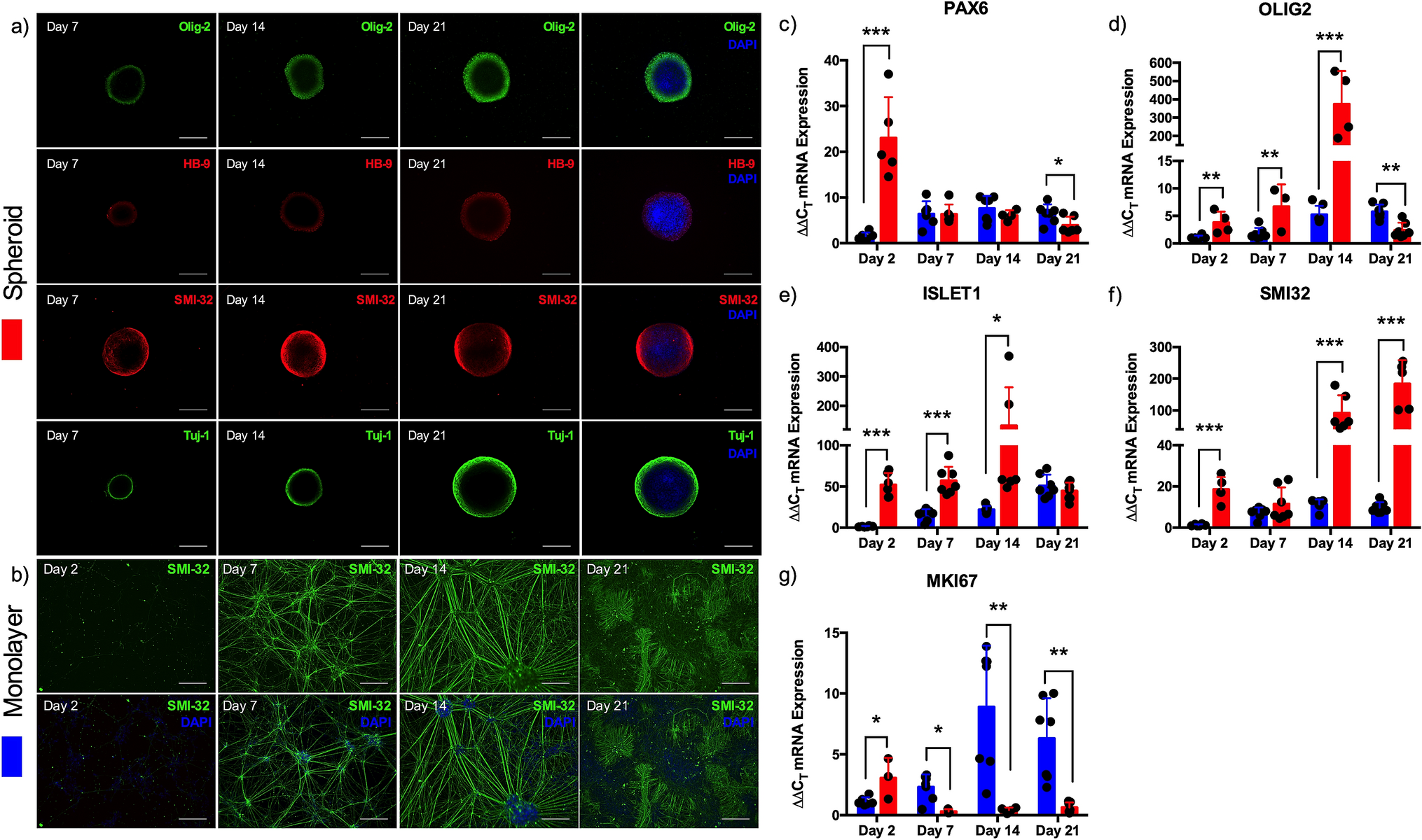
Identification and purity of SMNs by immunofluorescence with motor... |  Download Scientific Diagram
Identification and purity of SMNs by immunofluorescence with motor... | Download Scientific Diagram

SMI32 Antibody For Examining Axonal Damage Via IHC | Biocompare Antibody  Review
SMI32 Antibody For Examining Axonal Damage Via IHC | Biocompare Antibody Review

Bioengineered model of the human motor unit with physiologically functional  neuromuscular junctions | Scientific Reports
Bioengineered model of the human motor unit with physiologically functional neuromuscular junctions | Scientific Reports

Neuron Specific Nuclear Protein - an overview | ScienceDirect Topics

Abnormal SMI-31, SMI-34 and SMI-32 staining of Purkinje cells in MS... |  Download Scientific Diagram
Abnormal SMI-31, SMI-34 and SMI-32 staining of Purkinje cells in MS... | Download Scientific Diagram

Confocal images of cultured cells stained with CHAT (red) and SMI-32... |  Download Scientific Diagram
Confocal images of cultured cells stained with CHAT (red) and SMI-32... | Download Scientific Diagram

Abnormal SMI-31, SMI-34 and SMI-32 staining of Purkinje cells in MS... |  Download Scientific Diagram
Abnormal SMI-31, SMI-34 and SMI-32 staining of Purkinje cells in MS... | Download Scientific Diagram
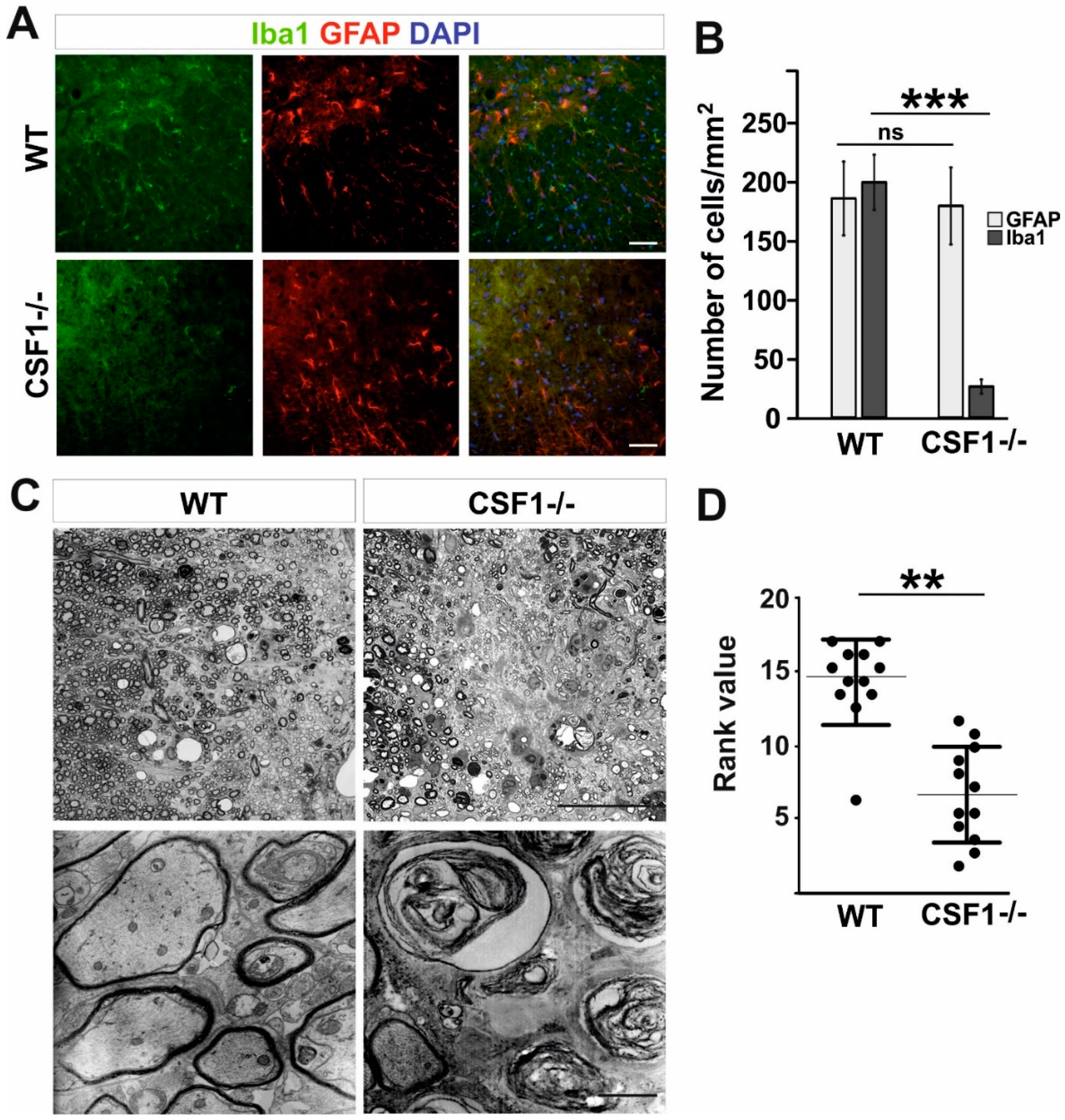
Cells | Free Full-Text | Csf1 Deficiency Dysregulates Glial Responses to  Demyelination and Disturbs CNS White Matter Remyelination
Cells | Free Full-Text | Csf1 Deficiency Dysregulates Glial Responses to Demyelination and Disturbs CNS White Matter Remyelination

SMI-32 + RGCs coexpress TRPV1 and TRPV4 signals. (A) A representative... |  Download Scientific Diagram
SMI-32 + RGCs coexpress TRPV1 and TRPV4 signals. (A) A representative... | Download Scientific Diagram

Distribution of SMI-32-stained elements inside the spinal cord. (A)... |  Download Scientific Diagram
Distribution of SMI-32-stained elements inside the spinal cord. (A)... | Download Scientific Diagram
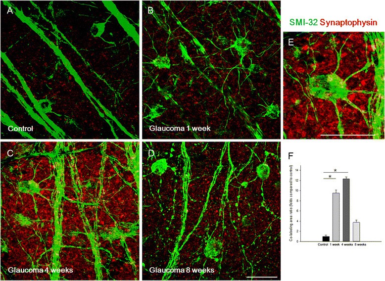
Alterations of the synapse of the inner retinal layers after chronic  intraocular pressure elevation in glaucoma animal model | Molecular Brain |  Full Text
Alterations of the synapse of the inner retinal layers after chronic intraocular pressure elevation in glaucoma animal model | Molecular Brain | Full Text

Axonal damage analysis. Observe the illustration of SMI-32 staining in... |  Download Scientific Diagram
Axonal damage analysis. Observe the illustration of SMI-32 staining in... | Download Scientific Diagram

The subpopulation-specific expression of SMI-32 and FNP-7... | Download  Scientific Diagram
The subpopulation-specific expression of SMI-32 and FNP-7... | Download Scientific Diagram

Alexa Fluor 594 anti-Neurofilament H NF-H Nonphosphorylated Antibody  anti-Neurofilament H - SMI 32
Alexa Fluor 594 anti-Neurofilament H NF-H Nonphosphorylated Antibody anti-Neurofilament H - SMI 32

Myelin Loss Does Not Lead to Axonal Degeneration in a Long-Lived Model of  Chronic Demyelination | Journal of Neuroscience
Myelin Loss Does Not Lead to Axonal Degeneration in a Long-Lived Model of Chronic Demyelination | Journal of Neuroscience